
After myocardial or vascular injury, repair is controlled by a timed interaction among innate and adaptive immune cells, endothelial cells, fibroblasts, extracellular matrix and surviving cardiomyocytes. Qaziim’s translational program measures these processes to define biological states and research hypotheses; it does not claim that a single biomarker can select patients for an unproven cell therapy.
Scientific interpretation framework
A clear framework helps visitors distinguish measurement, evidence and responsible translation into care or research.
Evidence level
Each finding is assessed against peer-reviewed literature, professional guidance, analytical validity and the maturity of clinical evidence. Research signals are identified explicitly and are not presented as established care.
Biological and clinical context
Molecular data are interpreted together with phenotype, family history, medicines, imaging, laboratory measurements and population context. No biomarker is meaningful in isolation.
Responsible output
Reports state the method, result, uncertainty, limitations and appropriate next step. Clinically relevant findings require qualified review; research models require validation before use in patient decisions.
Inflammation initiates repair
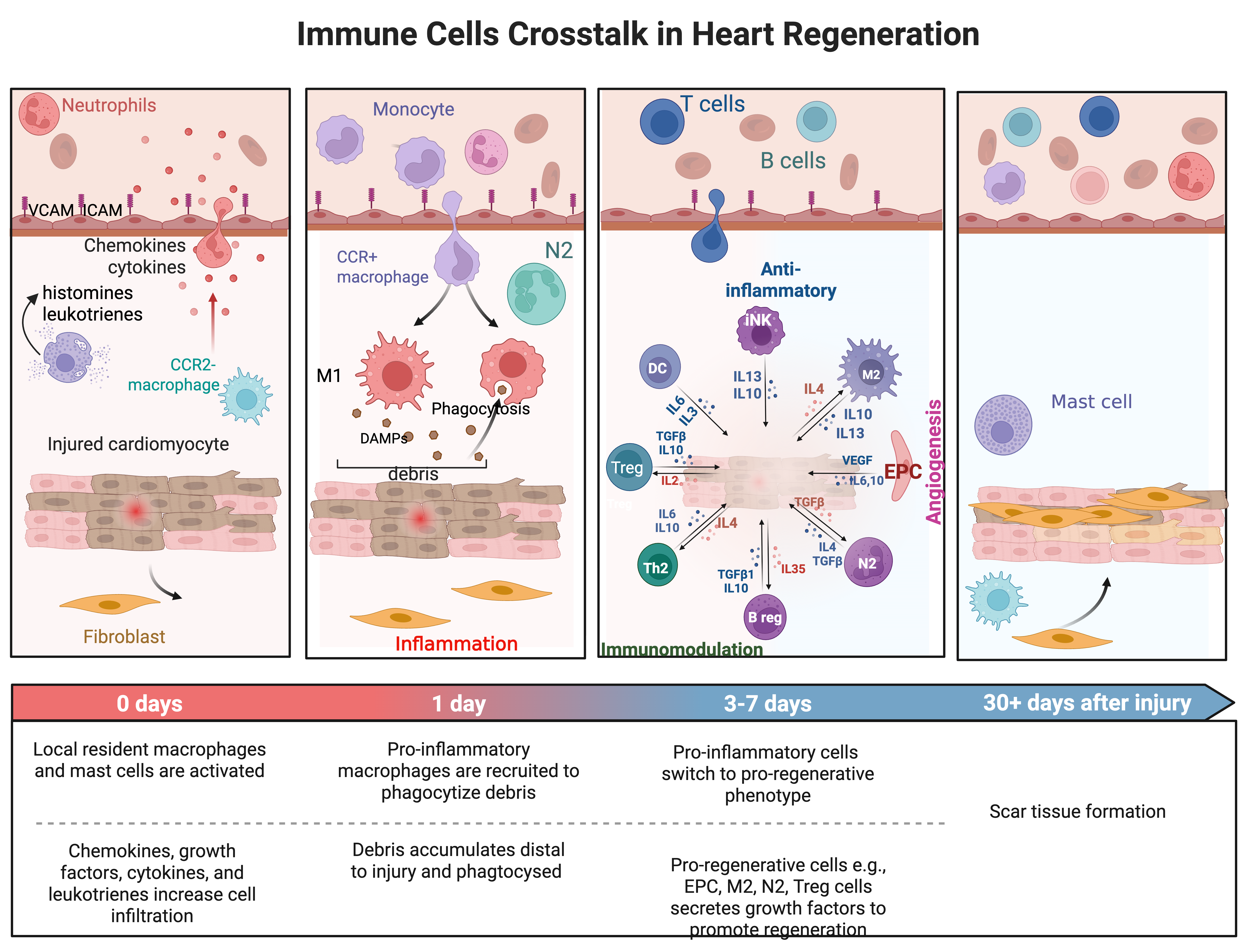
Injury releases damage-associated molecular patterns and chemokines. Neutrophils and inflammatory monocytes enter the tissue, remove debris and amplify cytokine signalling. This early response is necessary, but excessive or persistent inflammation can increase adverse remodelling.1
Resolution, fibrosis and remodelling
Macrophage phenotypes, regulatory lymphocytes and anti-inflammatory mediators then support resolution. Fibroblasts deposit extracellular matrix to preserve structural integrity; insufficient scar formation risks rupture, whereas excessive or persistent fibrosis can impair relaxation, conduction and ventricular function.2
Endothelial and vascular regeneration
Endothelial cells and endothelial progenitor-cell populations contribute to angiogenic signalling and vascular repair. Functional assays may examine migration, colony formation, nitric-oxide biology, oxidative stress and paracrine factors in diabetes, ischaemic heart disease and peripheral artery disease.3
Integrated regenerative research profile
Transcriptomics can describe active gene programs; proteomics and cytokines quantify signalling; cell-free DNA may reflect injury; imaging and clinical measures define tissue-level outcomes. Longitudinal sampling is essential because the same marker may have different meanings during inflammation, resolution and remodelling.3
Research design and meaningful endpoints
Mechanistic studies should use prespecified sampling times, standardized processing, appropriate control groups and clinically relevant endpoints such as ventricular function, perfusion, exercise capacity or validated patient-reported outcomes. Biomarker change alone does not establish therapeutic benefit.3
- Salybekov, A. A. et al. Therapeutic application of regeneration-associated cells. Stem Cell Res. Ther. 14, 203 (2023). doi:10.1186/s13287-023-03431-9
- Huang, H. & Huang, W. Regulation of endothelial progenitor cell functions in ischemic heart disease. Front. Cardiovasc. Med. 9, 896782 (2022). doi:10.3389/fcvm.2022.896782
- Frangogiannis, N. G. The inflammatory response in myocardial injury, repair, and remodelling. Nat. Rev. Cardiol. 11, 255–265 (2014). doi:10.1038/nrcardio.2014.28