
Regeneration-associated cells (RACs)
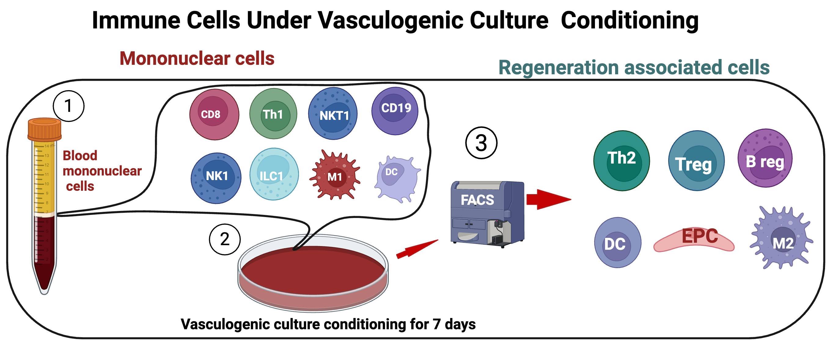
RAC is used in parts of the literature for selected or conditioned cell populations intended to support angiogenesis and immune–vascular coordination. The term is not a universally standardised cell identity; composition, processing and evidence must be stated for each product.1
In published experimental workflows, peripheral-blood mononuclear cells undergo vasculogenic culture conditioning. The resulting mixed population may be enriched for endothelial progenitor–associated cells and regulatory immune phenotypes. This is a manufacturing concept, not proof of clinical efficacy.
- Peripheral and critical limb ischaemia
- Diabetic vascular complications
- Myocardial ischaemia
- Chronic wounds
Endothelial progenitor–associated populations
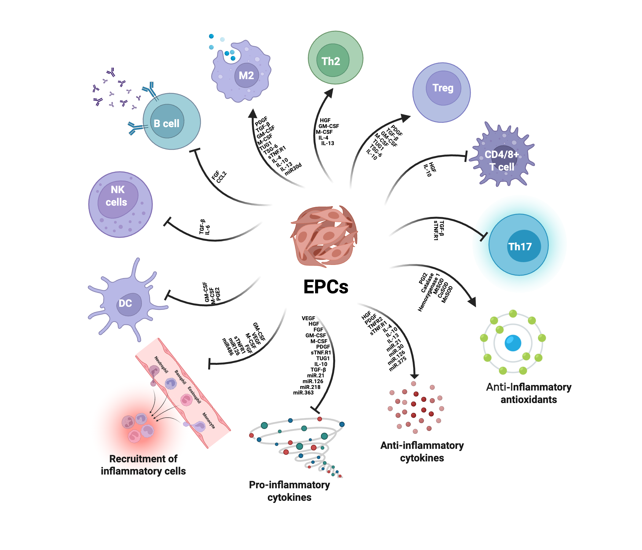
CD34 and CD133 enrich heterogeneous early haematopoietic or endothelial-associated progenitor populations. Investigated mechanisms include paracrine support, endothelial repair and neovascularisation rather than direct replacement of whole tissues.2
The EPC label has been used for biologically different populations. Rigorous reports therefore describe marker panels, culture conditions and functional assays instead of relying on one surface marker.
- Coronary and peripheral vascular disease
- Myocardial injury
- Neurovascular injury
- Tissue engineering
Mesenchymal stromal cells (MSCs)
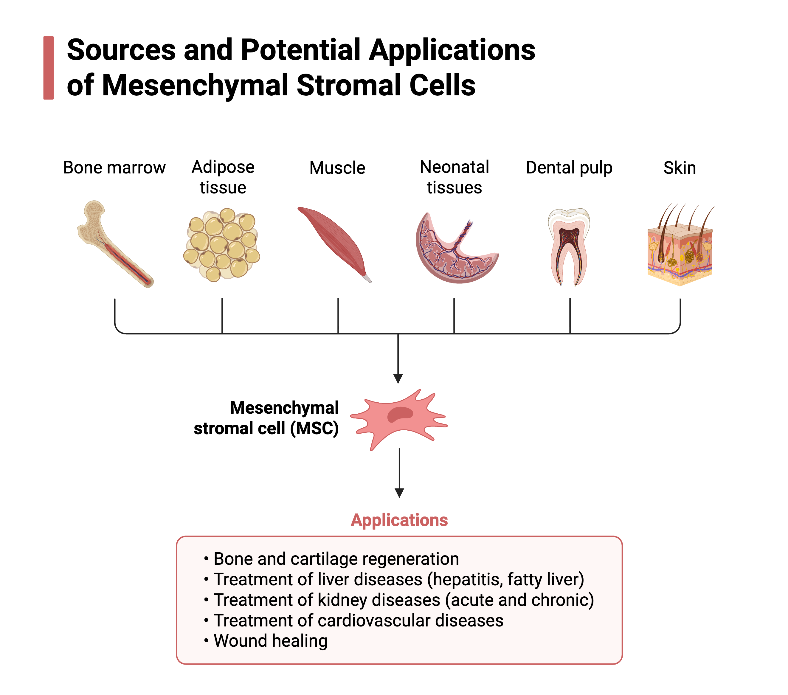
MSCs are extensively studied across inflammatory, vascular and musculoskeletal indications. Current models emphasise secreted cytokines, growth factors and extracellular vesicles, as well as context-dependent immunomodulation. Cell source, culture, dose and potency can materially change the product.3
MSCs can be isolated from several tissues, but cells from different sources are not automatically equivalent. Donor characteristics, passage number, oxygen conditions, cryopreservation and assay methods can alter phenotype and secretory activity.
- Osteoarthritis and connective-tissue injury
- Immune and inflammatory disease
- Kidney, liver and lung injury
- Cardiovascular and wound research
MSC differentiation and trophic activity
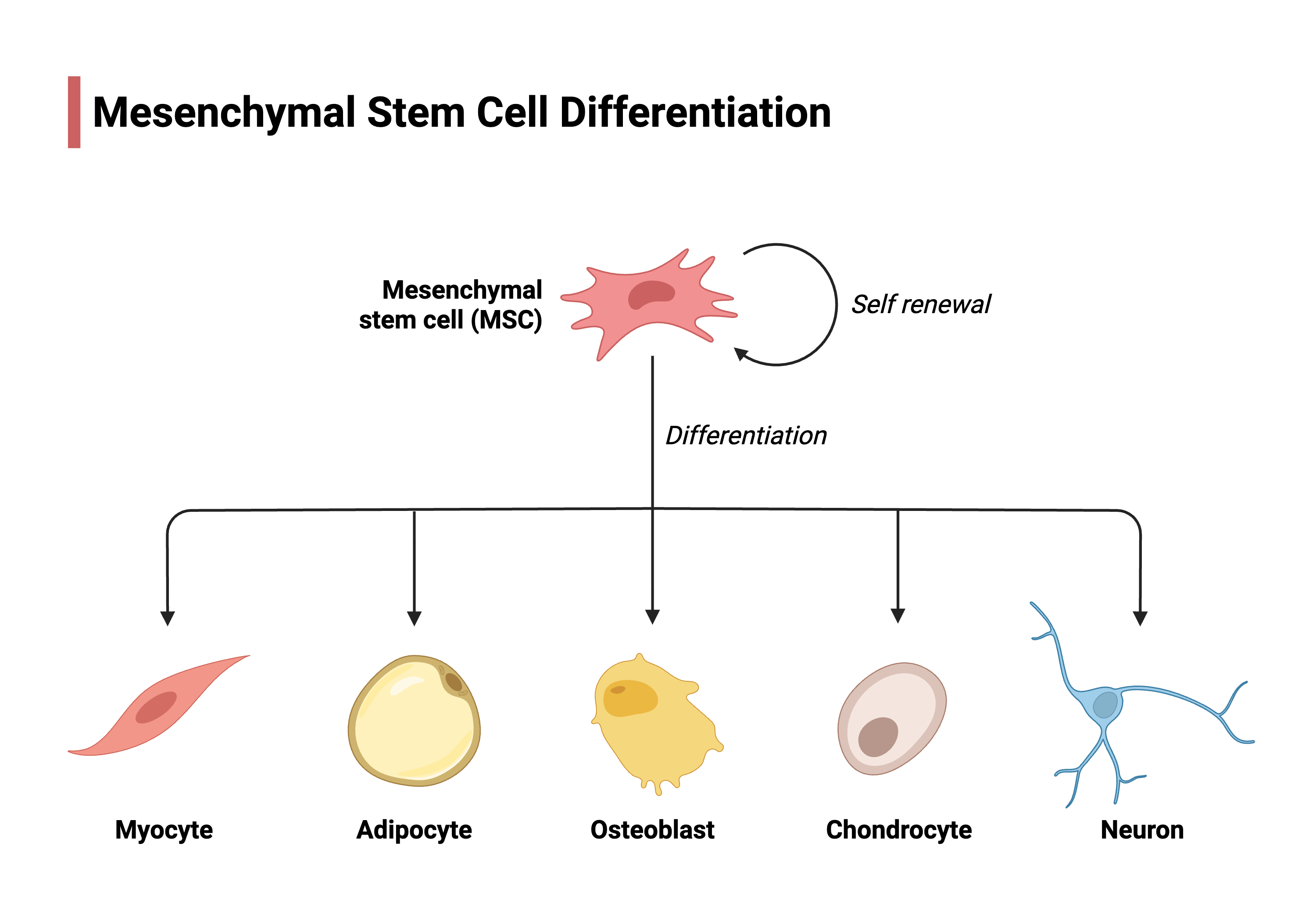
In defined laboratory conditions, MSC preparations can show osteogenic, chondrogenic and adipogenic differentiation. Claims of broad conversion into unrelated lineages require careful validation.4
For many proposed therapies, transient paracrine and immunomodulatory activity is considered more plausible than durable engraftment and replacement of damaged tissue.
Induced pluripotent stem cells (iPSCs)
Reprogrammed somatic cells can expand extensively and differentiate into many cell types. Established research uses include disease modelling, drug screening, organoids and gene-editing studies; clinical cell-replacement programmes require stringent purification and quality control.1
- Disease models and drug discovery
- Precision and gene-editing research
- Organoid and tissue engineering
- Experimental cell replacement
Extracellular vesicles (EVs)
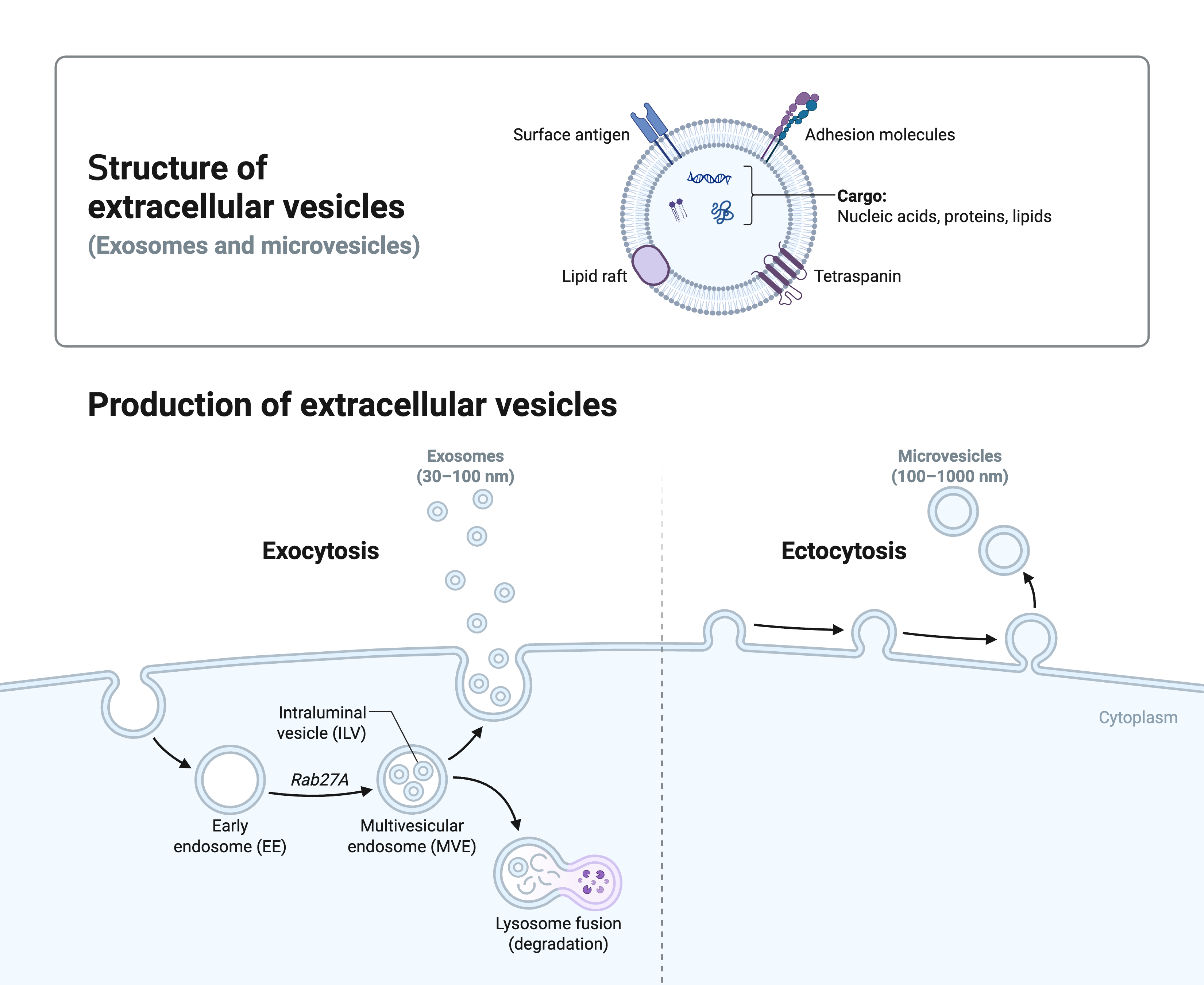
EVs are membrane-bound particles carrying proteins, RNA, lipids and metabolites between cells. They may mediate part of the paracrine activity attributed to transplanted cells, but therapeutic EV products still face challenges in identity, potency, dosing, scale-up and clinical validation.2
Small EVs may arise through the endosomal pathway, whereas larger vesicles can bud directly from the plasma membrane. Size ranges overlap, so current guidance recommends operational descriptions based on measured properties and preparation methods rather than assuming biogenesis from size alone.
- Cardiac, neurological and kidney injury
- Fibrosis and osteoarthritis
- Skin repair and diabetic wounds
- Immune-mediated disease
Product characterisation
Comparability, dose and delivery
The biological source, donor characteristics, isolation method, culture medium, passage, storage and thawing conditions can alter a cell product. A manufacturing change therefore requires comparability testing rather than an assumption that the old and new products are equivalent.3
Dose is not simply a cell count. Route of administration, viability at delivery, tissue retention, biodistribution and host immunity determine effective exposure. These variables explain why results cannot automatically be transferred between products that share a broad cell label.